Blank Diagram Of A Long Bone : Skeletal System Outline Printable Human Skeleton Diagram Labeled Unlabeled And Blank Skeleton Drawings Human Skeleton Human Skeletal System / Home » unlabelled » blank diagram of a long bone :
Blank Diagram Of A Long Bone : Skeletal System Outline Printable Human Skeleton Diagram Labeled Unlabeled And Blank Skeleton Drawings Human Skeleton Human Skeletal System / Home » unlabelled » blank diagram of a long bone :. Blank muscle diagram to label sketch coloring page. It can be found under the periosteum and in the diaphyses of long bones, where it provides support and protection. Layer of bone tissue that has many small spaces and is found j…. These include the bones of the arms and legs. Covers the surfaces of bones where they come together to form….
Choose from 500 different sets of long bone diagram flashcards on quizlet. Long bones have a thick outside layer of compact bone and an inner medullary cavity containing bone marrow. You need to get 100% to score the 10 points available. Smartdraw includes 1000s of professional healthcare and anatomy chart templates that you can modify and make your own. A long bone has two parts:
Free Anatomy Quiz The Anatomy Of Bones Quiz 1 from www.free-anatomy-quiz.com A long bone has two parts: The long bones have a long shaft and two bigger ends. Superficial muscles of the anterior trunk. Stability of the compact bone. Examples of long bones include the femur, tibia, fibula, metatarsals, and phalanges. The only short bones in the human skeleton are in the carpals of the wrists and the tarsals of the ankles. Skeletal system and long bone anatomy diagrams bundle. The diaphysis and the epiphysis.
Bone on side of the foot 12 photos of the bone on side of the foot bone on side of foot growing, bone on side of foot sticks out, fractured bone on side of foot, the bone on the side of my foot is sticking out, what is the bone on the side of my …
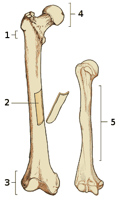
Most, but not all, features you are required to know are shown on the following pages. Layer of bone tissue that has many small spaces and is found j…. This is an online quiz called long bone diagram labeling. Label the parts of a long bone. It can be found under the periosteum and in the diaphyses of long bones, where it provides support and protection. Each system contains haversian canals surrounded by concentric. Bone on side of the foot 12 photos of the bone on side of the foot bone on side of foot growing, bone on side of foot sticks out, fractured bone on side of foot, the bone on the side of my foot is sticking out, what is the bone on the side of my … Parts of a long bone. Start studying long bone labeling. Related posts of long bone diagram labeled. The diaphysis and the epiphysis. Blank diagram of long bone. Hollow bone or long bone is longer than it is wide and is composed of the following elements image:
Long bone structure diagram and definitions flashcards quizlet. There is a printable worksheet available for download here so you can take the quiz with pen and paper. This is an online quiz called label the long bone. Long bones function as rigid bars that move when muscles contract. A whole skeletal muscle is considered an organ of the muscular system.each organ or muscle consists of skeletal muscle tissue, connective tissue, nerve tissue, and blood or vascular tissue.
Femur Bone Anatomy Labeled Diagram Quiz Color Coded Parts Skeletal System Lower Extremity Ezmed from images.squarespace-cdn.com End of a long bone. Long, short, flat, irregular and sesamoid. Several muscles that move the arms, head, and neck have their origins on the sternum. A whole skeletal muscle is considered an organ of the muscular system.each organ or muscle consists of skeletal muscle tissue, connective tissue, nerve tissue, and blood or vascular tissue. The diaphysis is the tubular shaft that runs between the proximal and distal ends of the bone. These include the bones of the arms and legs. Long bones are one of the five bone types that are classified by shape. Shaft of a long bone.
End of a long bone.
The structure of a long bone allows for the best visualization of all of the parts of a bone (figure 6.7). The diaphysis and the epiphysis. Each system contains haversian canals surrounded by concentric. The diaphysis and the epiphysis. The only short bones in the human skeleton are in the carpals of the wrists and the tarsals of the ankles. Most, but not all, features you are required to know are shown on the following pages. Body anatomy organs human muscle anatomy human skeleton anatomy anatomy bones gross anatomy anatomy and physiology quiz muscular system anatomy muscle diagram anatomy coloring book. It is 2 feet long and hollow, to make it lighter. Long bones have a thick outside layer of compact bone and an inner medullary cavity containing bone marrow. End of a long bone. A hollow medullary cavity is found in the center of long bones and serves as a storage area for bone marrow. Blank bone diagram rome fontanacountryinn com. Superficial muscles of the anterior trunk.
The epiphyseal line is a remnant of an area that contained hyaline cartilage that grew. A long bone has two parts: Long bones function as rigid bars that move when muscles contract. Shaft of a long bone. Smartdraw includes 1000s of professional healthcare and anatomy chart templates that you can modify and make your own.
The Bones Canadian Cancer Society from www.cancer.ca A = epiphysis b = diaphysis c = articular cartilage d = periosteum f = compact bone g = medullary cavity (yellow marrow) h = endosteum j = epiphyseal line (growth plate) coloring worksheet for this image. A long bone is a bone that has a shaft and 2 ends and is longer than it is wide. Choose from 500 different sets of long bone diagram flashcards on quizlet. Blank muscle diagram to label sketch coloring page. The largest bone in the body, the _____, is a long bone. It also protects several vital organs of the chest, such as the heart, aorta, vena cava, and. In long bones, as you move from the outer cortical compact bone to the inner medullary cavity, the bone transitions to spongy bone. Blank bone diagram rome fontanacountryinn com.
Blank muscle diagram to label sketch coloring page.
This is an online quiz called label the long bone. Examples of long bones include the femur, tibia, fibula, metatarsals, and phalanges. Hollow bone or long bone is longer than it is wide and is composed of the following elements image: The longest and the robust bone of the arm as observed in the following labeled diagram is called the humerus. Start studying long bone diagram. Long bones include the humerus (upper arm), radius (forearm), ulna (forearm), femur (thigh), fibula (thin bone of the lower leg), tibia (shin bone) , phalanges (digital bones in the hands and feet), metacarpals (long bones within the hand), and metatarsals (long bones. Long bones are hard, dense bones that provide strength, structure, and mobility. Smartdraw includes 1000s of professional healthcare and anatomy chart templates that you can modify and make your own. Most, but not all, features you are required to know are shown on the following pages. Choose from 500 different sets of flashcards about long bone diagram on quizlet. Image of a typical long bone is shown with numbers identifying the various parts, such as the epiphysis. The long bones have a long shaft and two bigger ends. The structure of a long bone allows for the best visualization of all of the parts of a bone (figure 6.7).


0 Komentar